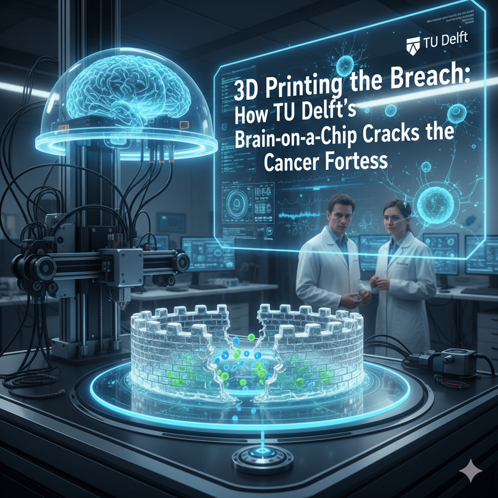
TU Delft researchers use advanced 3D bioprinting to create "brain-on-a-chip" models, simulating how treatments can finally breach the "fortress" of complex cancer tumors
A team at TU Delft has 3D-printed a functional human blood-brain-tumour barrier using two-photon polymerisation and microfluidics. The result is a research tool that can observe how glioma cells breach the barrier at molecular resolution — something no animal model has been able to provide. Here is what the science shows, what it does not yet show, and why the regulatory context makes the timing significant.
Analysis drawing on the published TU Delft research, FDA Modernization Act 2.0 documentation, EMCDDA organ-on-a-chip market data, and peer-reviewed literature on translational pharmacology · Updated March 2026
The blood-brain barrier is one of the most consequential obstacles in modern medicine. It is the reason that more than 98% of small-molecule drugs and nearly all large-molecule therapeutics fail to reach the brain in therapeutically meaningful concentrations. It is why glioblastoma — the most common and most lethal primary brain tumour in adults — has a median survival of approximately 15 months despite decades of intensive research investment. And it is why understanding exactly how tumour cells manipulate the barrier to their advantage has been so difficult: until recently, the only ways to study the barrier in a dynamic, living context were animal models that replicate it imperfectly, or post-mortem human tissue that captures a static endpoint rather than a process.
The work published by Associate Professor Angelo Accardo and his team at TU Delft’s Department of Precision and Microsystems Engineering represents a meaningful advance on both limitations. Using two-photon polymerisation to print a three-dimensional scaffold at sub-micron resolution, then seeding it with human endothelial cells, pericytes, and glioma cells under controlled microfluidic flow, the team produced a blood-brain-tumour barrier (BBTB) model that exhibits the structural and functional characteristics of the human barrier — including the tight junction integrity and flow-responsive behaviour that animal and flat-culture models have historically failed to replicate together.
What they can now observe, in real time, is the molecular sequence by which glioma cells interact with and breach that barrier. That observation is the research output. It is significant. It is also the beginning of a research programme, not the conclusion of one.
The Technical Achievement: Why Two-Photon Polymerisation Matters
Standard bioprinting approaches — extrusion-based or inkjet methods — produce scaffolds at resolutions of approximately 100–200 micrometres. The structural features that govern cell behaviour at the blood-brain barrier operate at scales of 1–10 micrometres: the geometry of the basement membrane, the spacing of tight junction proteins, the curvature of capillary walls that influences how endothelial cells orient and polarise.
Two-photon polymerisation (2PP) achieves resolution below one micrometre by using a pulsed near-infrared laser to initiate photopolymerisation only at the precise focal point where two photons are simultaneously absorbed — a nonlinear optical process that confines the reaction to a volume smaller than the diffraction limit of conventional light. The result is a scaffold that can be printed with feature sizes and surface geometries that cells respond to as they would to native extracellular matrix architecture.
This matters because cell behaviour is not purely chemical. Endothelial cells forming the blood-brain barrier require specific geometric and mechanical cues to form functional tight junctions — the protein complexes that give the barrier its characteristic impermeability. On flat 2D surfaces, these junctions form poorly. In 3D scaffolds with insufficiently precise geometry, they form inconsistently. The TU Delft approach addresses both problems by providing a substrate that accurately replicates the physical environment in which these cells normally operate.
The microfluidic integration adds the second critical dimension: fluid flow. The blood-brain barrier in vivo is continuously subjected to shear stress from blood flow, and this mechanical signal is one of the primary regulators of tight junction formation and maintenance. Static culture systems — including many existing 3D models — miss this entirely. The TU Delft model applies controlled flow through the printed vascular channel, producing a barrier that forms and behaves under conditions that more closely approximate the in vivo environment.
The Tri-Culture Model: Why Cell Composition Matters
Earlier blood-brain barrier models typically used monocultures — a single cell type, usually human cerebral microvascular endothelial cells (hCMEC/D3 is the most widely used line), seeded onto a membrane. These models produce measurable barrier function but consistently underestimate barrier tightness relative to in vivo measurements, because the endothelial cells lack the signals they normally receive from neighbouring cell types.
The TU Delft model uses a tri-culture system incorporating three cell populations that together constitute the neurovascular unit:
Endothelial cells form the vascular wall and are the primary structural component of the barrier. Their tight junctions determine permeability.
Pericytes wrap around the endothelial cells and regulate barrier tightness through direct contact signalling and paracrine communication. Research published in Nature Neuroscience has shown that pericyte loss is one of the earliest observable changes in blood-brain barrier breakdown in neurodegenerative disease.
Glioma cells are included in the BBTB (blood-brain-tumour barrier) variant specifically to model the tumour microenvironment. Glioblastoma cells are known to secrete factors that alter barrier permeability in ways that facilitate tumour invasion and, paradoxically, also impede drug delivery. Including them in the model allows observation of these interactions directly.
The combination produces a barrier with transendothelial electrical resistance (TEER) values — the standard measure of tight junction integrity — that are substantially higher than monoculture equivalents and closer to in vivo measurements. This is the metric that determines whether a model is actually useful for drug permeability testing: a barrier that is too leaky will overestimate how much drug reaches the brain, invalidating the prediction.
What This Enables: The Research Applications
The immediate research application is mechanistic: the model allows observation of the molecular events involved in glioma invasion of the barrier at a resolution and in a dynamic context that was not previously achievable. Specifically, the team has used it to observe the sequence of cell-cell signalling events — the “molecular handshake” between tumour cells and endothelial cells — that precedes barrier disruption. Understanding this sequence identifies potential intervention points that could either prevent invasion or exploit the disruption pathway to improve drug delivery.
The second application is pharmacological screening. A functional BBTB model with validated permeability characteristics can be used to test candidate drug compounds for their ability to cross the barrier before committing to animal studies or clinical trials. This does not replace later-stage testing — it provides an earlier filter that reduces the number of compounds that progress to animal studies with no realistic prospect of CNS penetration.
The third, longer-term application is personalised medicine. The model can in principle be constructed using cells derived from a specific patient’s tumour biopsy, creating a patient-specific BBTB that reflects that individual’s unique tumour microenvironment. Drug screening on this personalised model could inform treatment selection before systemic administration — a “clinical trial of one” conducted on a chip rather than in a patient. This application remains at the proof-of-concept stage; the cell sourcing, preparation, and quality control requirements for routine clinical use are substantially more demanding than for research use.
The Regulatory Context: FDA Modernization Act 2.0
The TU Delft breakthrough arrives at a moment of significant regulatory change that materially affects its commercial and clinical relevance. The FDA Modernization Act 2.0, signed into US law in December 2022, removed the previous statutory requirement that drug sponsors conduct animal testing before submitting an Investigational New Drug (IND) application. It explicitly authorises the FDA to accept data from “microphysiological systems” — which includes organ-on-a-chip models — as part of the preclinical evidence package.
This is a necessary but not sufficient condition for adoption. The Act removes the legal barrier to using MPS data; it does not create a validated pathway for how that data is interpreted and weighted relative to traditional preclinical packages. The FDA’s Center for Drug Evaluation and Research (CDER) and the National Centre for the Advancement of Translational Sciences (NCATS) are both actively developing qualification frameworks for specific organ-on-a-chip platforms — the process by which a specific model is validated for a specific intended use and its data is given defined evidentiary weight in regulatory submissions.
The European Medicines Agency (EMA) operates a parallel qualification process for novel methodologies. Both processes are methodical and time-consuming by design: regulators need to understand a platform’s performance characteristics, failure modes, and inter-laboratory reproducibility before they can define how its data should be used in safety and efficacy assessments.
The practical implication for the TU Delft model is that regulatory qualification for use in drug submissions is a multi-year process from current proof-of-concept. The research value is immediate. The commercial and regulatory impact will take longer to realise.
The “Animal Model Era” Framing: What the Evidence Supports
Claims that animal models are being superseded or that a new era is beginning deserve some precision, because the actual picture is more nuanced than either the boosters or the sceptics typically acknowledge.
Animal models have genuine and documented limitations for CNS drug development. A 2022 analysis in PLOS Biology examining 101 CNS drug candidates that showed efficacy in rodent models found that the majority failed in Phase II human trials, with species differences in receptor pharmacology, blood-brain barrier composition, and neuroinflammatory pathways cited as contributing factors. The translational problem is real.
At the same time, animal models provide systemic context that no current organ-on-a-chip system can replicate: the interaction between organ systems, immune surveillance, pharmacokinetic distribution across tissues, and the chronic toxicity profiles that emerge over weeks or months of exposure. A chip that accurately models one barrier or one organ cannot observe what happens when a drug reaches the liver, is metabolised, and the metabolite reaches the kidney. These systemic questions will require animal data or human clinical data for the foreseeable future.
The more accurate framing — supported by the National Institutes of Health’s strategic plan for organ-on-a-chip development and the NCATS Tissue Chip for Drug Screening programme — is that MPS platforms are being developed as complements to animal testing that can reduce the number of animals used, improve the predictive value of preclinical data, and filter out compounds that will fail for human-specific reasons before they reach animal or clinical stages. Complete replacement of animal models in drug development is a long-term aspiration rather than a near-term reality.
The organ-on-a-chip market is projected to grow substantially through the decade, with estimates ranging from $500 million to over $1 billion by 2030 depending on the scope of the analysis. The growth is driven by pharmaceutical company adoption of these platforms for early-stage screening — not by regulatory mandates requiring their use, which do not yet exist in this form.
The Neurodegenerative Disease Opportunity
The application area where the translational failure of animal models is perhaps most acutely felt — and where the TU Delft model’s capabilities are most directly relevant — is neurodegenerative disease.
Alzheimer’s disease has produced more Phase III clinical trial failures than any other therapeutic area: over 200 failed trials between 2002 and 2022, representing more than $40 billion in research investment. Many of these failures involved compounds that showed clear efficacy in mouse models of amyloid pathology and then demonstrated no clinical benefit in human trials. The Alzheimer’s Association has supported investment in human-relevant models specifically because the mouse amyloid models have proven poor predictors of human disease course.
A functional human blood-brain barrier model that can be used to test whether compounds cross the barrier in relevant concentrations — and to observe how the barrier itself changes under conditions that mimic early Alzheimer’s pathology — addresses one specific but important component of this failure pattern. It does not address the question of whether amyloid clearance is the correct therapeutic target, or whether the compounds tested have off-target effects in other tissues. But it provides a human-relevant filter at a stage where the current pipeline has no equivalent.
Parkinson’s disease presents a similar pattern: dopaminergic neuron models in rodents have guided drug development that has consistently underperformed in clinical translation, partly because the rodent blood-brain barrier has different transporter expression profiles than the human barrier for many of the compounds being tested.
Conclusion: A Research Tool With Significant but Bounded Implications
The TU Delft blood-brain-tumour barrier model is a genuine technical achievement. The combination of two-photon polymerisation scaffolding, tri-culture cell composition, and microfluidic integration produces a model that addresses specific, documented limitations of existing approaches — particularly the failure of earlier models to form tight junctions under physiologically realistic conditions and the inability to observe glioma-barrier interactions dynamically.
Its implications for drug discovery are real but require accurate framing. It is a research tool that enables mechanistic observations not previously possible and an early-stage screening platform that can improve the predictive value of preclinical CNS drug development. It is not a replacement for the full preclinical and clinical development pipeline, and the regulatory qualification process required before its data can formally substitute for animal data in drug submissions is a multi-year process from where it currently stands.
The broader shift it represents — toward human-relevant, mechanistically accurate in vitro models as a primary component of early drug discovery — is genuine and accelerating. The FDA Modernization Act 2.0, the NCATS tissue chip programme, and the growing pharmaceutical industry investment in MPS platforms all point in the same direction.
What the TU Delft work adds to that direction is a specific, validated demonstration that the blood-brain barrier — historically one of the most difficult human barriers to model accurately — can now be printed, populated with human cells, and used to observe disease mechanisms in real time. For the millions of patients with glioblastoma, Alzheimer’s disease, and Parkinson’s disease for whom the translational gap has meant decade after decade of promising preclinical results that did not survive contact with human biology, that demonstration is worth taking seriously.
Sources & Further Reading
- TU Delft — Angelo Accardo research group, Precision and Microsystems Engineering
- FDA Modernization Act 2.0 — FDA overview and guidance
- NCATS Tissue Chip for Drug Screening programme
- European Medicines Agency — Qualification of novel methodologies
- Nature Neuroscience — Pericyte loss and blood-brain barrier breakdown
- Nature Biomedical Engineering — Two-photon polymerisation in tissue engineering
- Alzheimer’s Association — Clinical trials and translational research
- Michael J. Fox Foundation — Parkinson’s disease research and translational challenges
- Grand View Research — Organ-on-a-chip market forecast
- PLOS Biology — CNS drug translational failure analysis
- Emulate Bio — Commercial organ-on-a-chip platforms
- Mimetas — Organ-on-a-chip for BBB modelling